Most people visit the optometrist for blurry vision, new glasses, or dry eyes. But what many don’t realize is that a routine eye exam can do far more — it can literally save lives.
That’s because the eye is the only place in the body where doctors can directly observe blood vessels, nerves, and brain-connected tissue without surgery. And in some cases, it’s an optometrist who first spots the warning signs of serious neurological diseases.
Let’s explore how the eye provides a window into the brain — and the critical role optometrists play in early diagnosis of life-threatening conditions.
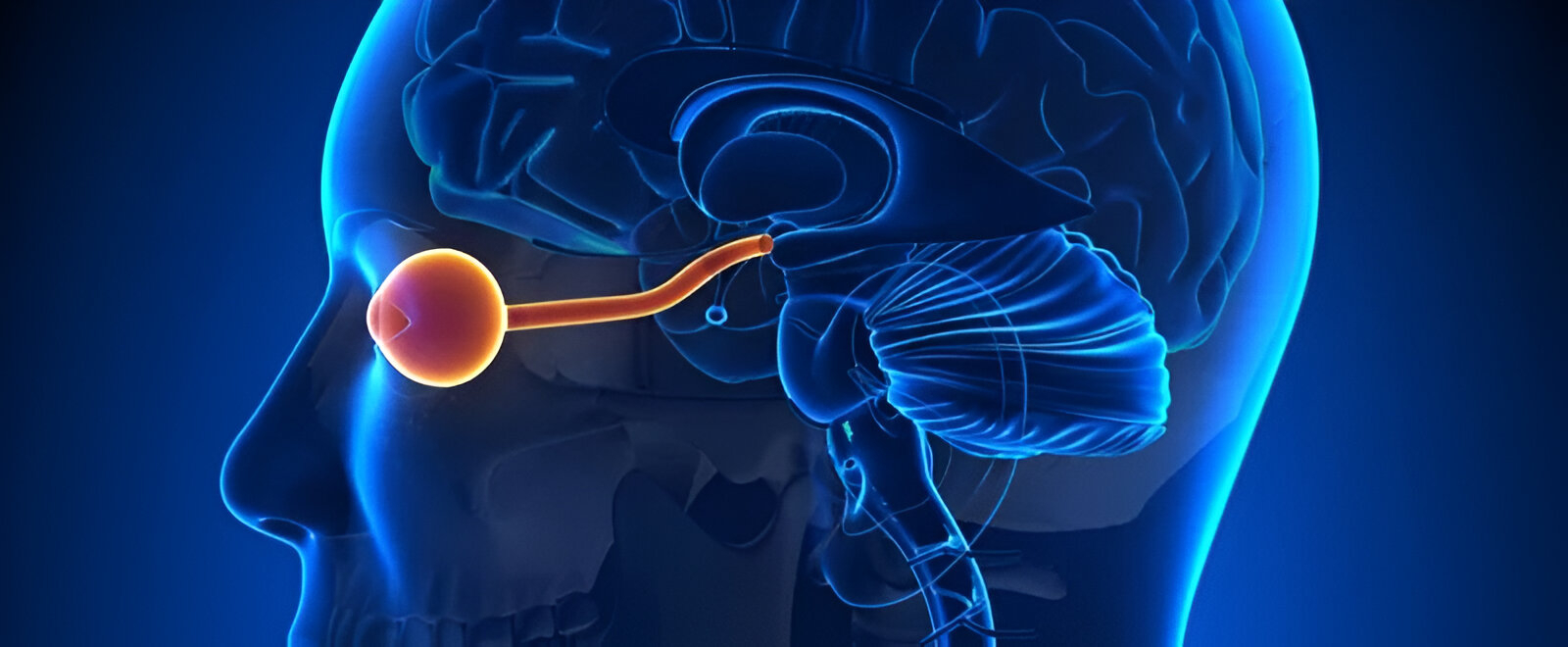
The Optic Nerve: A Direct Brain Connection
The optic nerve is actually an extension of the brain. It transmits visual information from the retina to the brain’s visual cortex. Since it’s made of central nervous system tissue, any disease affecting the brain or nerves can often show up here first.
“The eye is the only part of the brain you can see directly.”
— Dr. Andrew Lee, Neuro-Ophthalmologist
Conditions First Detected During an Eye Exam
Here are some major neurological conditions that optometrists may identify during a routine exam:
1. Multiple Sclerosis (MS)
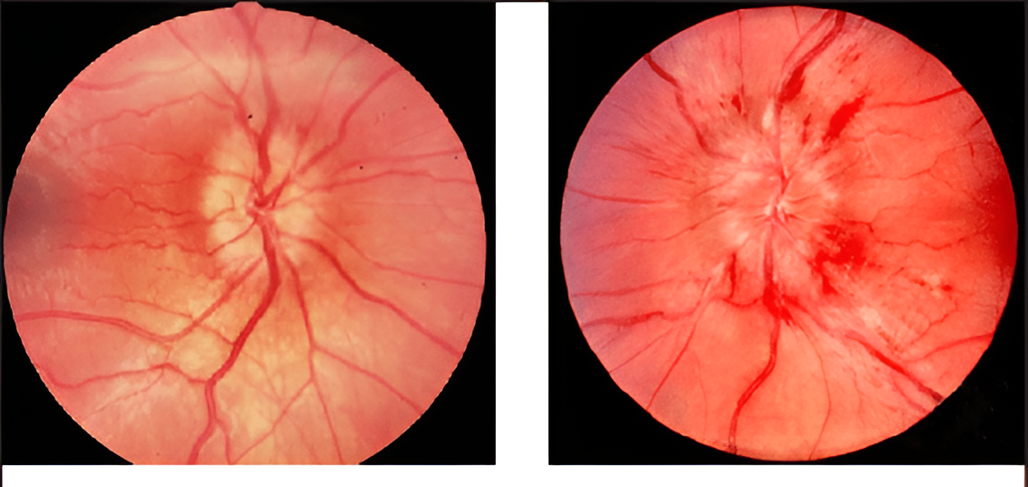
One of the first signs of MS is optic neuritis — inflammation of the optic nerve.
-
Patients may complain of sudden vision loss, pain with eye movement, or blurred vision.
-
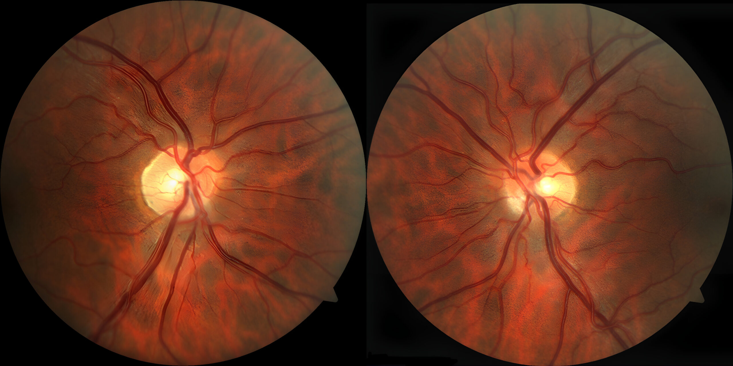
Fundus exams or OCT imaging may show optic disc swelling or later, optic atrophy.
-
Optometrists may refer the patient for neurological testing and MRI, helping diagnose MS early.
Fun fact: About 20% of MS patients experience visual symptoms as their first sign.
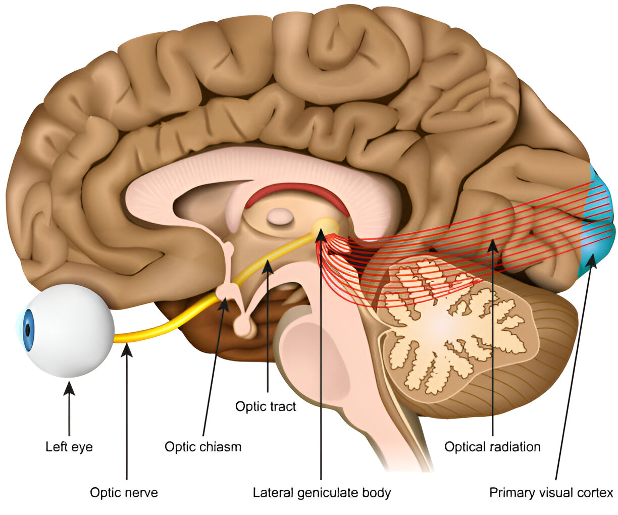
2. Brain Tumors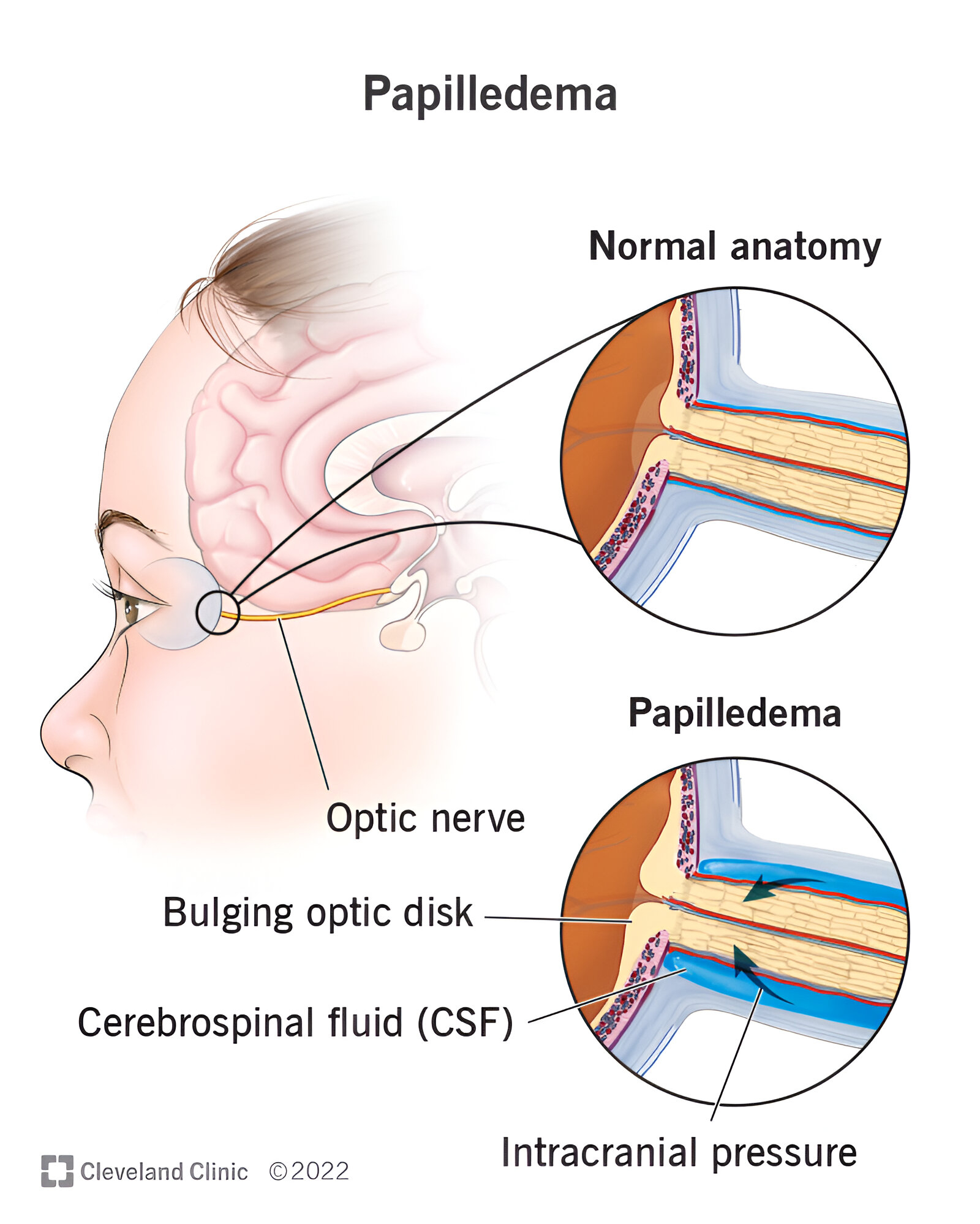
Tumors near the optic chiasm or occipital lobe can affect visual pathways.
-
Signs include visual field defects (like tunnel vision or blind spots).
-
Optometrists may perform a visual field test, spotting suspicious patterns.
-
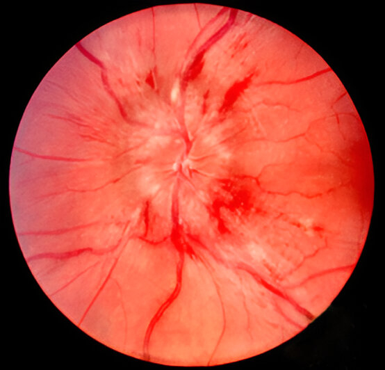
Papilledema (swollen optic nerves) may also be seen in fundus exams if the tumor increases intracranial pressure.
3. Idiopathic Intracranial Hypertension (IIH)
A condition where pressure builds up in the skull without a tumor or lesion.
-
Seen mostly in young women with obesity.
-
Symptoms: Headache, transient visual obscurations, double vision.
-
Optometrists can detect papilledema or sixth nerve palsy causing diplopia.
Early detection can prevent permanent vision loss.
4. Stroke or Transient Ischemic Attack (TIA)
Sudden loss of vision in one eye, visual field defects, or eye movement abnormalities may indicate a vascular event in the brain.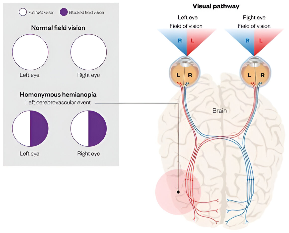
-
Optometrists may identify homonymous hemianopia or other patterns suggesting post-chiasmal stroke.
-
Timely referral for neuroimaging can be life-saving.
5. Myasthenia Gravis
This autoimmune condition causes muscle weakness, often first noticeable in the eyes:
-
Ptosis (drooping eyelid)
-
Diplopia (double vision)
-
Fluctuating symptoms worsening with fatigue
Optometrists may perform simple fatigue tests or ice pack tests and refer to neurologists for further workup.
6. Aneurysms or Vascular Malformations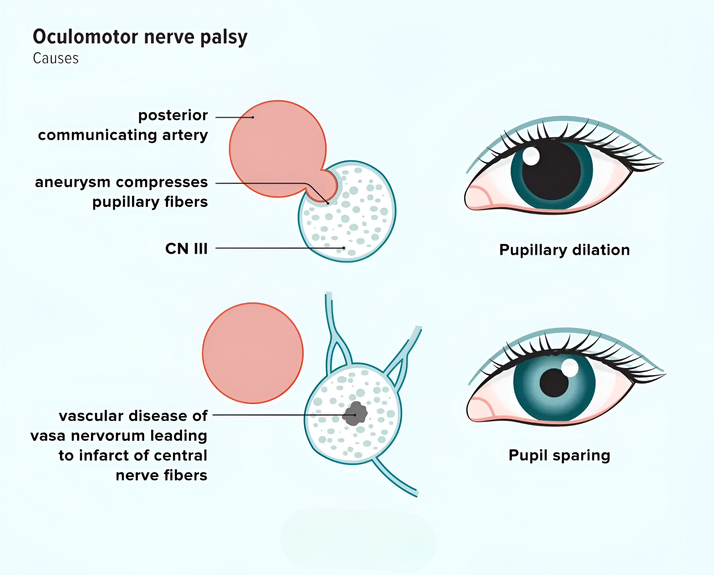
An aneurysm pressing on the oculomotor nerve can cause:
-
Ptosis
-
Dilated pupil
-
Eye movement problems
These are red-flag findings that require urgent brain imaging.
Tools Optometrists Use to Detect Brain Problems
Modern optometry is far more than just eye charts and lenses. Here’s what helps optometrists connect eye findings to the brain:
-
OCT (Optical Coherence Tomography) – high-resolution imaging of the optic nerve and retina.
-
Visual field testing (Perimetry) – detects subtle vision loss patterns linked to brain damage.
-
Fundoscopy – reveals swelling, bleeding, or changes in the optic disc.
-
Pupil reflex testing – abnormalities can indicate nerve pathway issues.
-
Eye movement tests – detect nerve palsies or coordination issues.
Why This Matters: Optometry as the Frontline of Brain Health
In many places — especially in countries like India — optometrists are the first point of contact for vision-related complaints. That places them in a critical position to screen for neurological red flags and make life-saving referrals.
With increased integration under health authorities like NCAHP (National Commission for Allied and Healthcare Professions), optometrists in India are becoming even more empowered to play a neuro-ophthalmic screening role.
Real Patient Example
A 25-year-old woman visits an optometrist with complaints of mild blurring and headache. A fundus exam reveals bilateral papilledema.
She’s urgently referred for a brain MRI — revealing benign intracranial hypertension. Early intervention prevents permanent vision loss.
Final Thoughts
The next time you go for an eye check-up, remember: your optometrist isn’t just checking how well you see. They might be the first to catch a brain tumor, multiple sclerosis, or stroke warning sign.
The eyes may truly be the windows to the soul — but they’re also diagnostic windows into the brain.
References
-
Lee, A.G., & Brazis, P.W. (2020). Clinical Pathways in Neuro-Ophthalmology.
-
American Academy of Ophthalmology – “The Eye and the Brain.”
-
Optometry Times – “How Optometrists Detect Neurological Conditions.”
-
National Institute of Neurological Disorders and Stroke (NINDS)
-
Reddy et al. (2021). “Role of Optometrists in Neuro-Visual Health.” Indian Journal of Optometry.
