
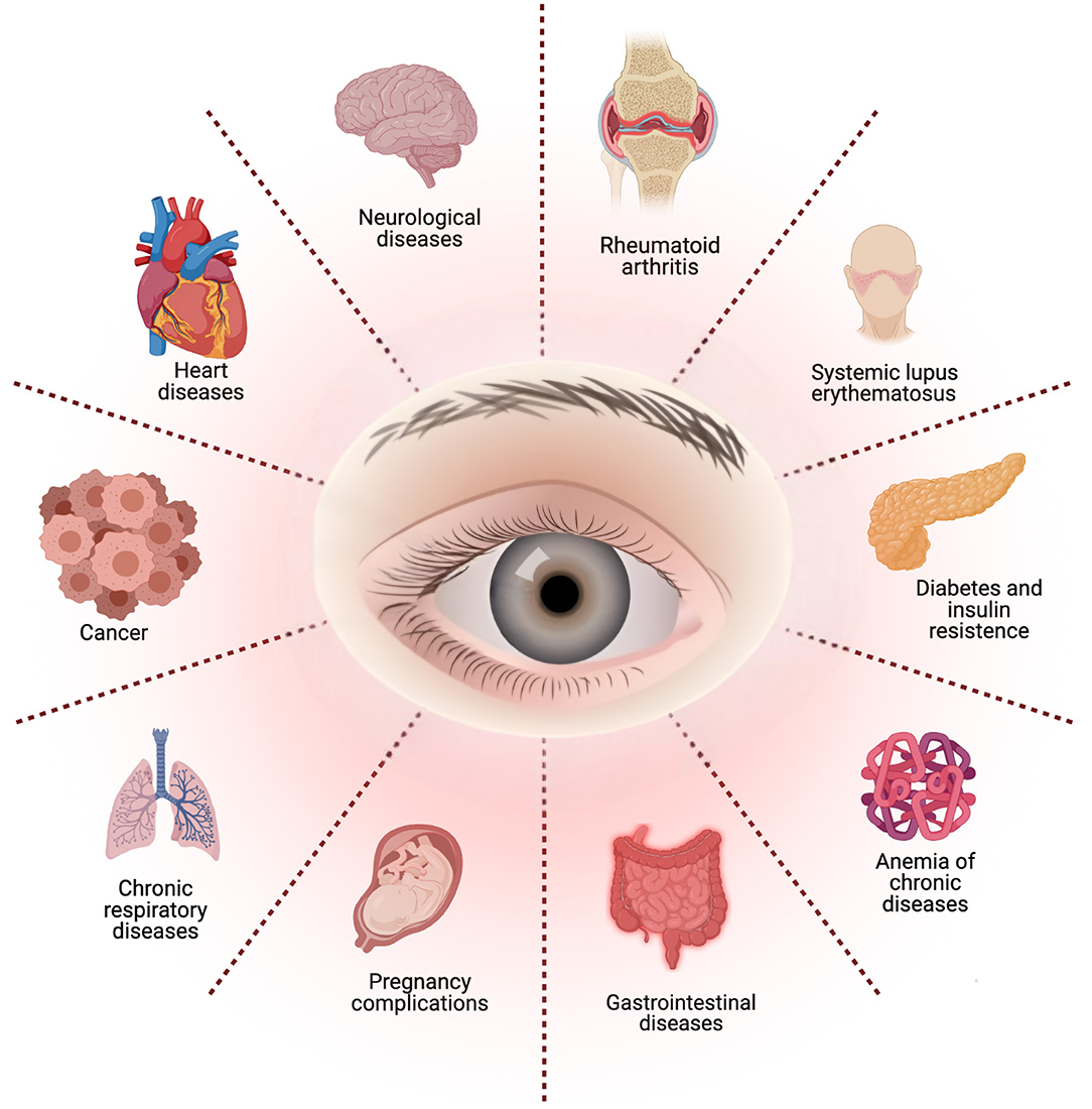
The Eyes Reveal More Than Just Vision Problems
The eyes are often called the windows to the soul—but in the world of medicine, they’re more accurately described as windows to overall health. A comprehensive eye exam can reveal not just issues with your eyesight, but also clues to serious systemic diseases—often before other symptoms appear.
As optometrists, our role extends beyond visual correction. In many cases, we’re the first healthcare professionals to detect signs of diabetes, hypertension, autoimmune diseases, and even brain tumors.
Let’s explore how your eye exam can uncover what’s happening throughout your entire body.
Why the Eye Is So Revealing
The eye is unique in that it allows direct, non-invasive visualization of blood vessels, nerves, and connective tissue. With tools like ophthalmoscopy, OCT (Optical Coherence Tomography), and fundus photography, we can inspect the retina, optic nerve, and blood vessels in high detail—without needing to cut open the body.
This makes the eye an ideal place to detect early signs of systemic disease.
Systemic Diseases That Show Up in the Eyes
Diabetes Mellitus → Diabetic Retinopathy

Observation:
- In early stages, we may see microaneurysms (small bulges in blood vessels), dot-and-blot hemorrhages, and hard exudates (lipid deposits).
- As the disease progresses, neovascularization—abnormal new vessel growth—may occur, which can lead to vitreous hemorrhage, tractional retinal detachment, and severe vision loss.
Clinical insight:
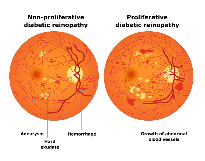
Diabetic retinopathy is classified into non-proliferative and proliferative stages. It can remain asymptomatic for a long time, making annual eye exams essential for diabetic patients—even those with good blood sugar control. Macular edema, in particular, is a leading cause of central vision loss and may require anti-VEGF injections or laser treatment.
Importance:
Ophthalmic signs may precede systemic symptoms, especially in patients with undiagnosed or poorly controlled diabetes. Detecting retinopathy can motivate patients to adhere to medication, diet, and glucose monitoring—making the eye exam a pivotal moment for early intervention and disease management.
Eye exams are essential even for asymptomatic diabetics—retinal changes can appear before noticeable vision loss.
Hypertension → Hypertensive Retinopathy
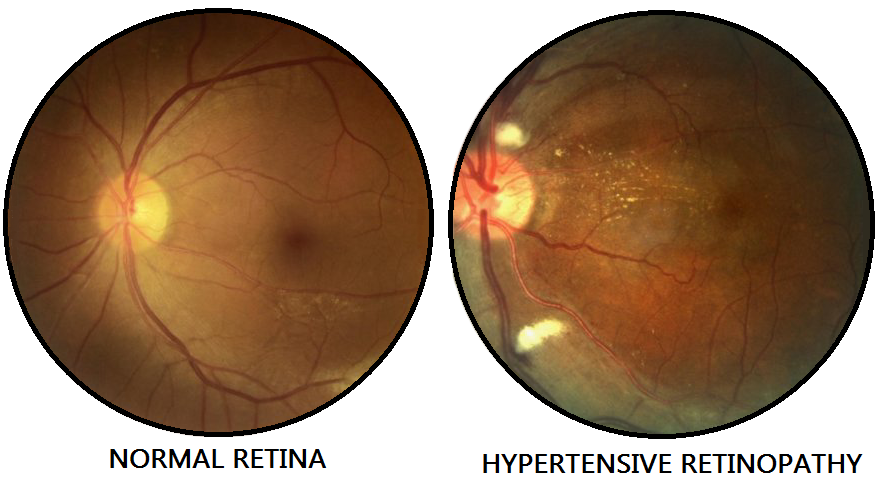
Observation:
Elevated blood pressure causes vascular changes like arteriolar narrowing, AV nicking (compression of venules by arteriosclerotic arterioles), cotton wool spots (nerve fiber infarcts), and flame-shaped hemorrhages.
In severe cases, optic disc edema may indicate malignant hypertension.
Clinical insight:
The Keith-Wagener-Barker classification system helps assess the severity of hypertensive retinopathy. Advanced signs indicate long-standing or uncontrolled hypertension and significantly increase the risk of stroke, heart attack, and kidney failure.
Impotance:
Eye findings can prompt referral to a cardiologist or internist. Sometimes, optometrists are the first to uncover silent hypertension—especially in patients who are asymptomatic or non-compliant with treatment.
Some patients first learn they have hypertension from their eye exam!
Autoimmune Diseases → Scleritis, Uveitis, Dry Eye
Observation: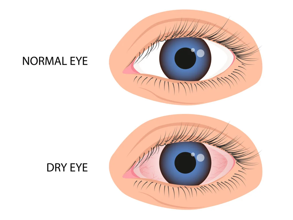
Autoimmune disorders such as Rheumatoid Arthritis, Systemic Lupus Erythematosus, or Ankylosing Spondylitis may present with episcleritis, scleritis (severe pain, redness, and photophobia), or anterior uveitis (inflammation of the iris and ciliary body).
Chronic inflammation also contributes to severe dry eye, especially in Sjögren’s Syndrome.
Clinical insight:
These patients may not have a systemic diagnosis yet. The presence of bilateral scleritis or recurrent uveitis can be a red flag that prompts systemic workup, including ANA, RF, HLA-B27, or ACE tests.
Importance:
Eye symptoms often correlate with systemic disease activity. Collaborative management with rheumatologists ensures proper immunosuppressive therapy and monitoring of ocular complications like posterior synechiae, cataract, or glaucoma.
Severe dry eye could be a sign of Sjögren’s syndrome, often missed in general medicine.
Multiple Sclerosis → Optic Neuritis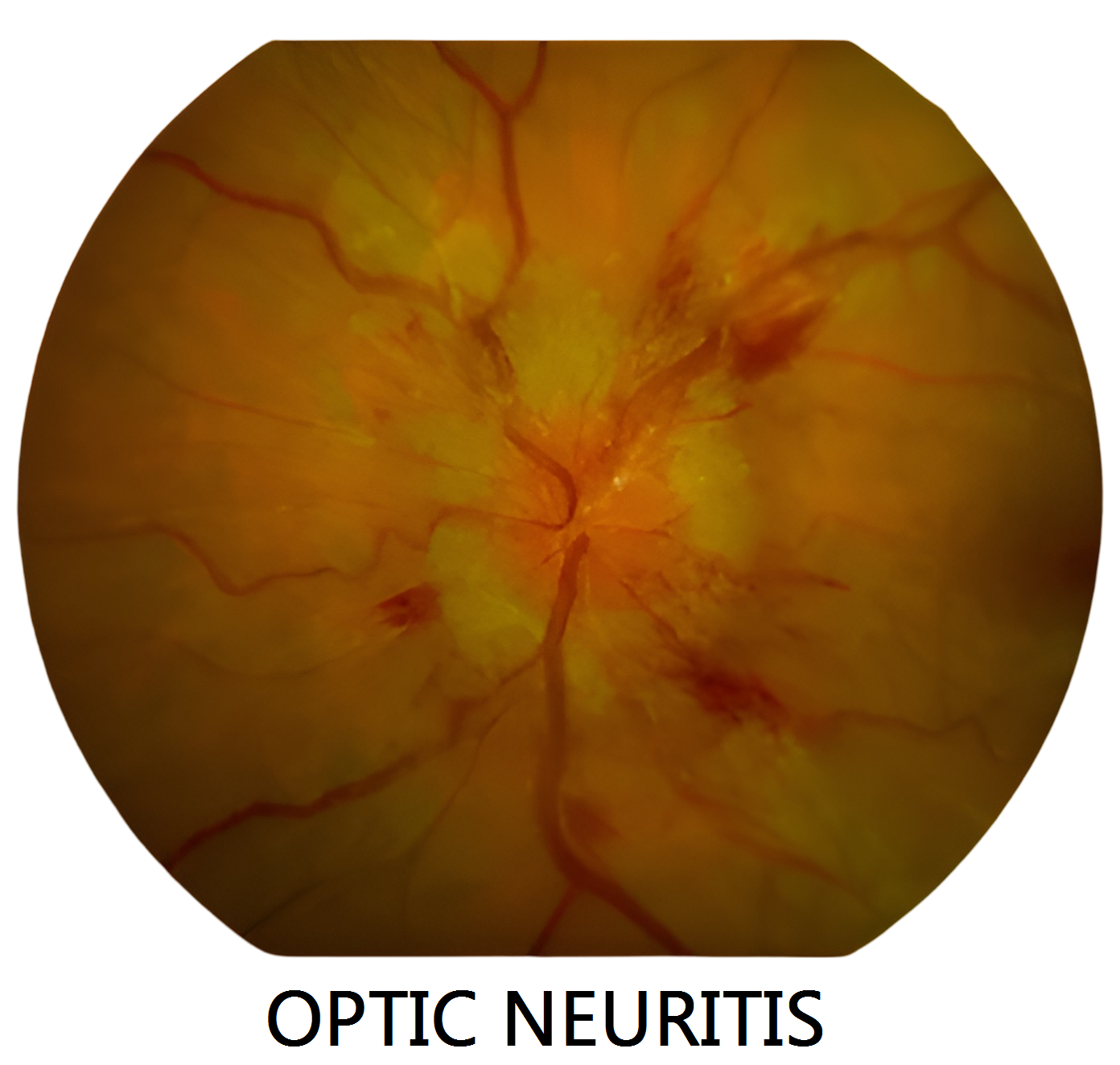
Observation:
Patients may report sudden vision loss, dull pain with eye movement, and color desaturation.
The optic disc may appear swollen (papillitis) or normal (retrobulbar neuritis). Visual field testing often reveals central scotomas.
Clinical insight:
Up to 50% of MS patients experience optic neuritis, and it is the initial presentation in approximately 20% of cases. MRI of the brain and orbits typically shows demyelinating plaques. Visual evoked potentials (VEPs) can assist in diagnosis.
Importance:
Early diagnosis allows timely initiation of disease-modifying therapy, which may reduce relapse rates and slow disease progression. Patients may also benefit from visual rehabilitation and counseling about prognosis.
Up to 25% of MS patients experience optic neuritis as the first symptom.
Thyroid Disease (Graves’) → Thyroid Eye Disease (TED)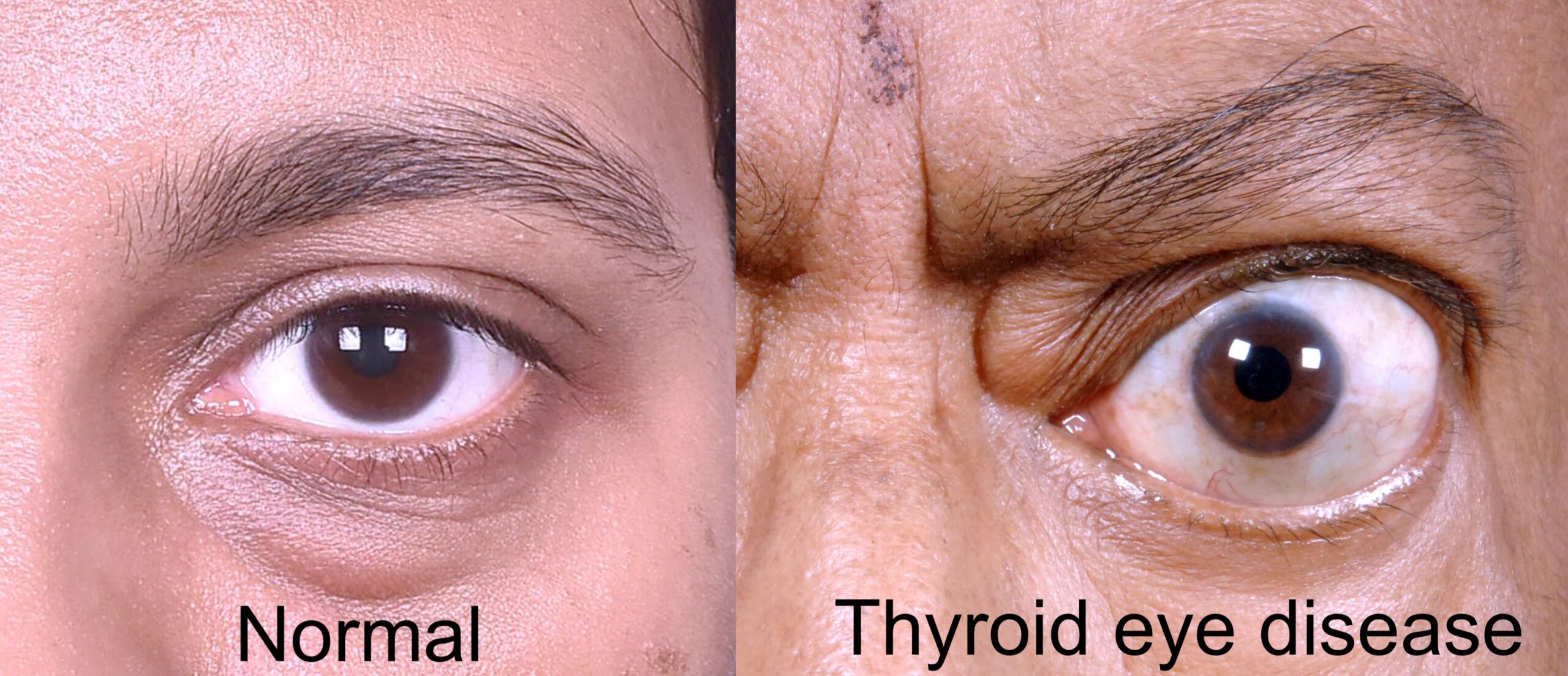
Observation:
Classic signs include lid lag, lid retraction, exophthalmos (proptosis), diplopia, and exposure-related dry eye.
Severe cases can involve optic nerve compression and vision loss.
Clinical insight:
TED occurs due to autoimmune inflammation of extraocular muscles and orbital fat. It may worsen after radioactive iodine therapy and is strongly associated with smoking. Orbital imaging (CT or MRI) helps assess severity.
Importance:
Ocular changes may occur before or even without thyroid dysfunction, especially in euthyroid Graves’ disease. Identifying early TED helps initiate protective strategies such as lubrication, smoking cessation, or referral for orbital decompression.
Patients often seek help for “bulging eyes” or cosmetic changes—leading to the discovery of an overactive thyroid.
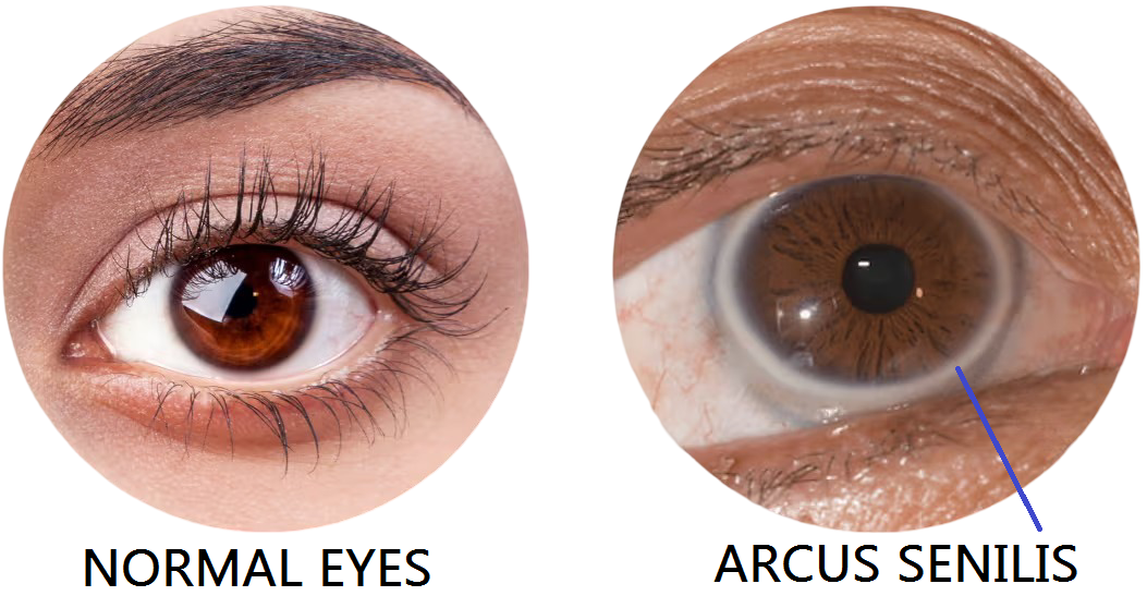
High Cholesterol → Hollenhorst Plaques & Arcus Senilis
Observation:
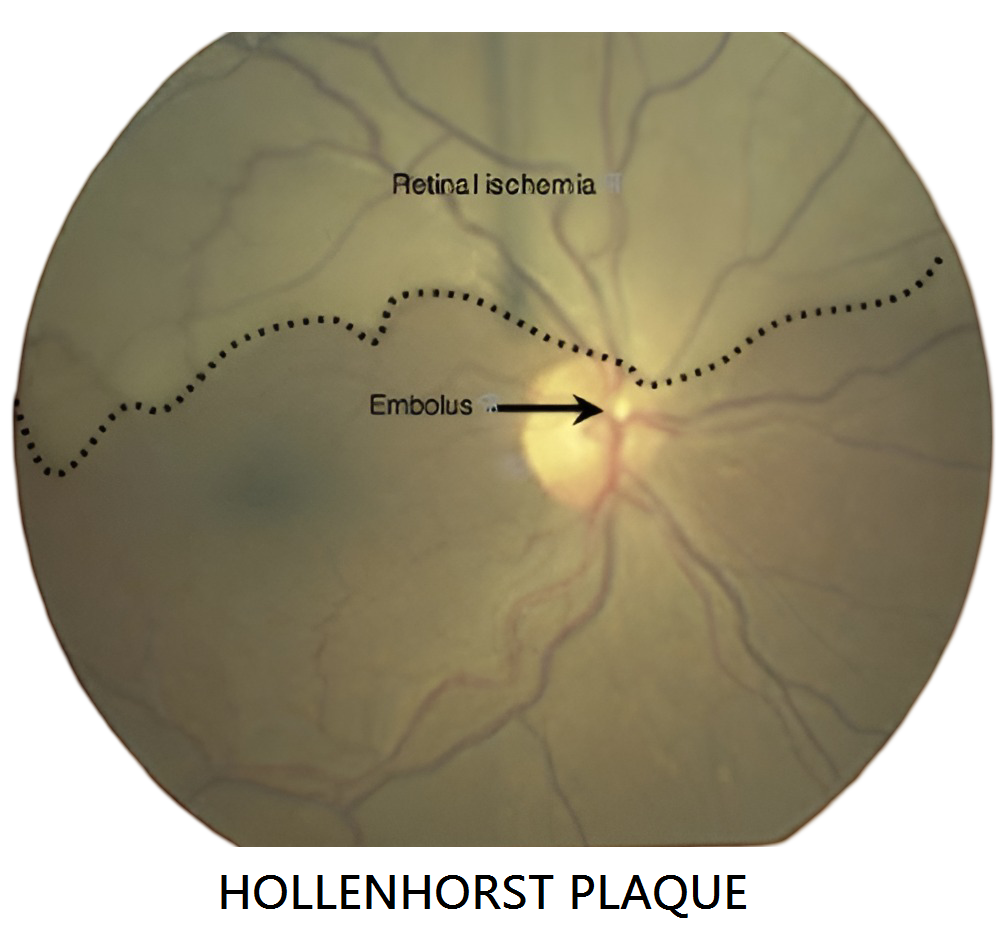
-
Hollenhorst plaques appear as bright, refractile emboli lodged in retinal arteries—usually at bifurcations.
-
Arcus senilis presents as a grayish-white ring at the corneal periphery, often benign in elderly but concerning in younger patients.
Clinical insight:
A retinal embolus may signal carotid artery disease or cardiac valve disease. Carotid Doppler ultrasound and lipid profile testing are typically indicated. Arcus under age 40 should trigger a full lipid panel.
Importance:
Early detection of emboli can prevent catastrophic events like stroke or myocardial infarction. Eye findings prompt life-saving cardiovascular referrals.
An eye exam can literally help prevent a heart attack or stroke.
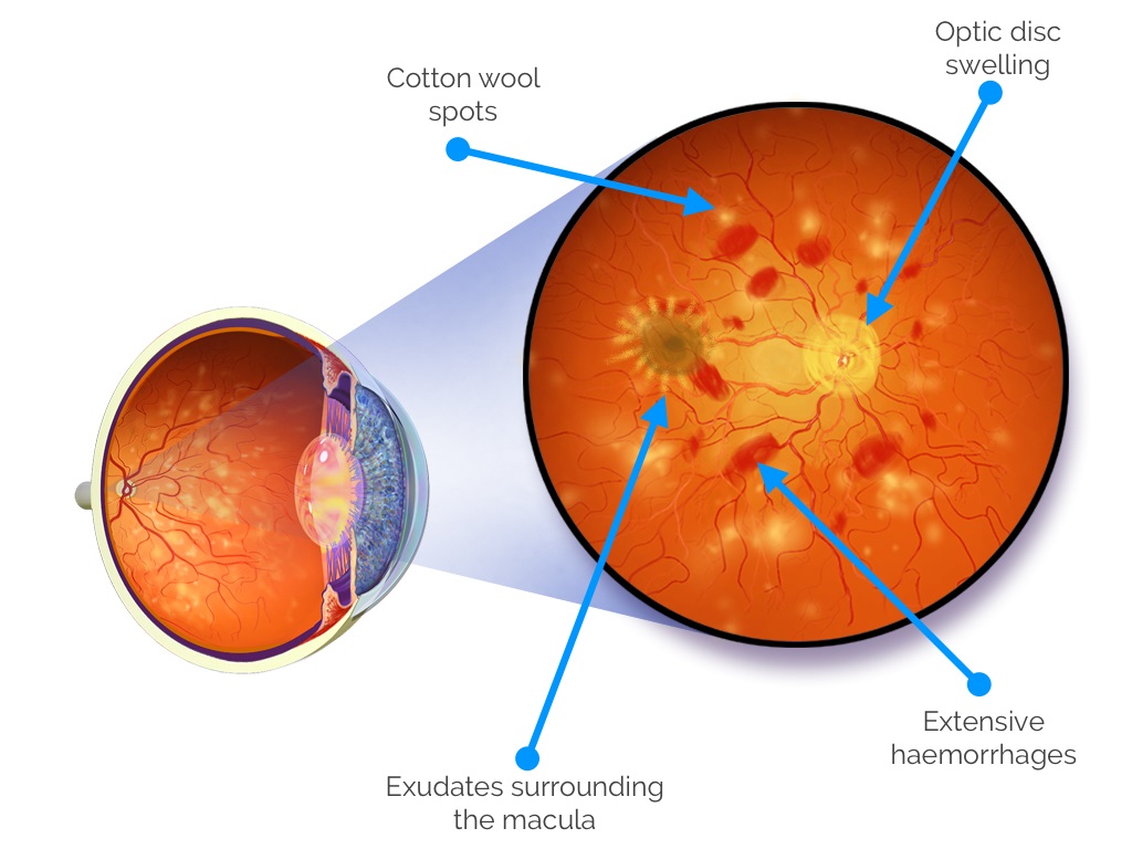
Brain Tumors → Papilledema & Cranial Nerve Palsies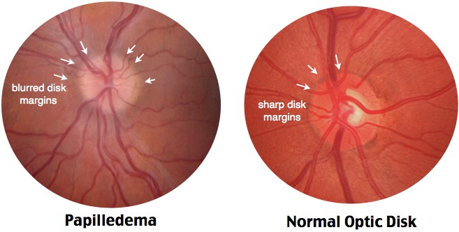
Observation:
Increased intracranial pressure can cause papilledema—bilateral optic disc swelling, often with blurred margins, hemorrhages, and engorged vessels. Sixth nerve palsy may present as horizontal diplopia. Visual field defects and headaches are common.
Clinical insight:
Papilledema is a medical emergency. MRI or CT imaging is necessary to rule out space-occupying lesions, hydrocephalus, or idiopathic intracranial hypertension (especially in young obese women).
Importance:
An eye exam could lead to early tumor detection, preventing long-term neurologic damage or death. Some patients present to the optometrist with only visual symptoms before a brain tumor is diagnosed.
“Silent” brain tumors can first reveal themselves through double vision or vision loss.
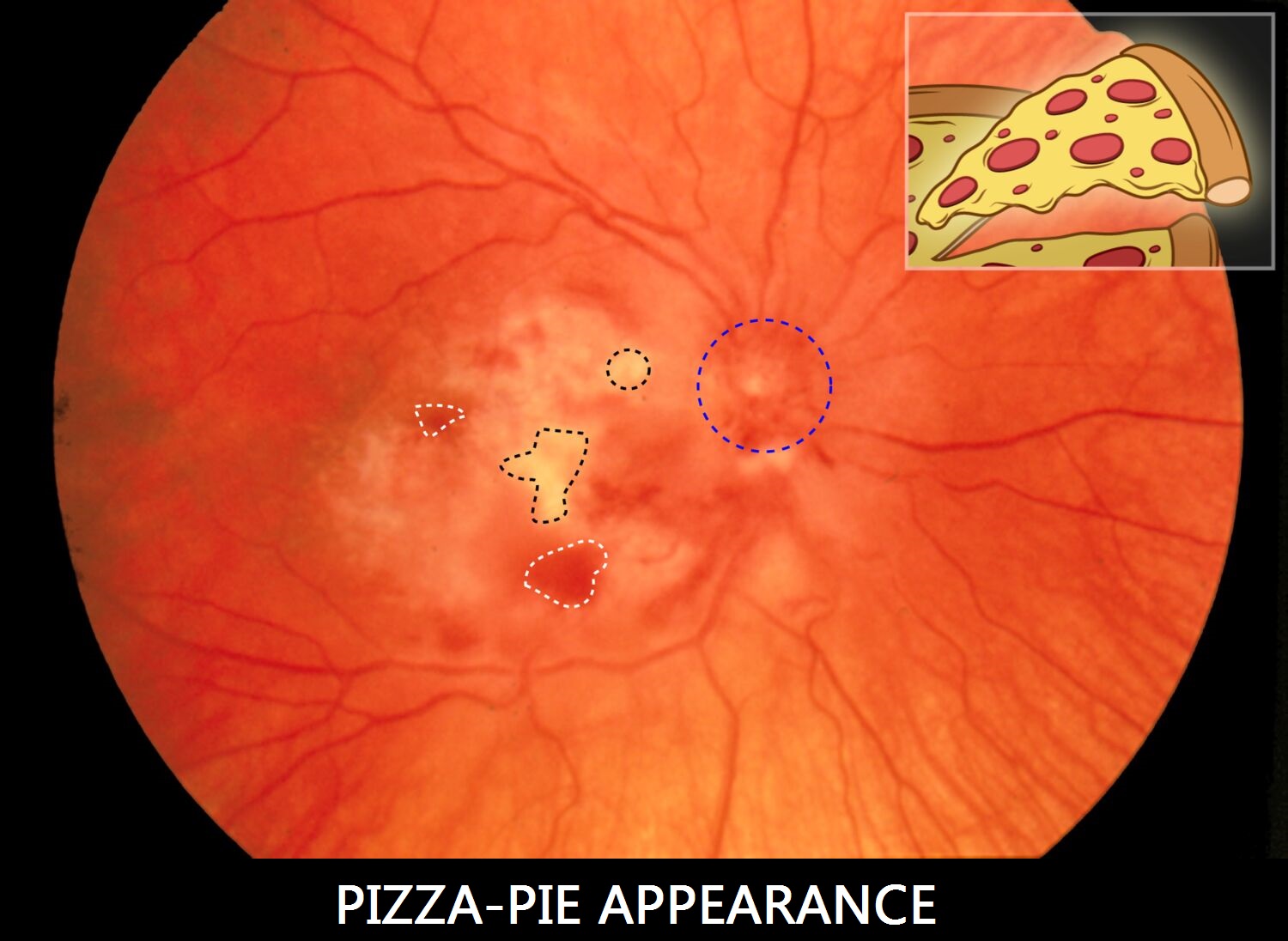
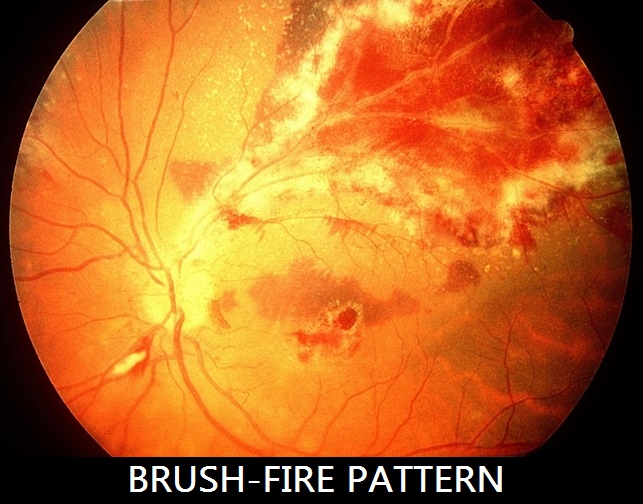
Infectious Diseases → HIV, Syphilis, TB, CMV Retinitis
Observation:
HIV-positive patients may show cotton wool spots, retinal hemorrhages, or cytomegalovirus (CMV) retinitis, which appears as “pizza pie” or brushfire patterns of necrosis.
Syphilis can cause posterior uveitis, interstitial keratitis, and retinal vasculitis.
Clinical insight:
Ocular manifestations may reflect immune status. The eye may be the first location of clinical symptoms, especially with ocular syphilis, which has been resurging in many regions. Lab tests like RPR, VDRL, HIV ELISA, and TB Quantiferon aid diagnosis.
Importance:
Timely identification can reduce vision loss and systemic spread. It also triggers appropriate testing and counseling for infectious disease management and partner notification.
Ocular syphilis has been on the rise globally—often diagnosed first by eye care professionals.
Tools That Help Detect Systemic Disease
| Tool | Use |
|---|---|
| Fundus Photography | High-res images of retina and optic nerve |
| OCT | Microscopic cross-sections of retina/nerve layers |
| Visual Field Testing | Detects neurological damage or tumors |
| Tonometry | Assesses intraocular pressure (glaucoma, tumors) |
| Fluorescein Angiography | Evaluates blood flow in retina (diabetes, vasculitis) |
The Optometrist’s Expanding Role
Modern optometrists are not just refraction experts—we’re frontline healthcare providers. Many patients visit their optometrist more regularly than their primary doctor. This makes the eye exam a crucial opportunity for:
-
Early detection of systemic disease
-
Timely referrals to specialists
-
Patient education and prevention
Final Thoughts
The eyes may be small, but they hold immense diagnostic power. Whether it’s spotting a dangerous spike in blood pressure, catching early signs of diabetes, or detecting neurological conditions—the eye exam is a window into the health of the whole body.
So the next time someone asks, “Why do I need an eye exam if my vision is fine?” — tell them:
It’s not just about seeing clearly. It’s about seeing the whole picture.
